

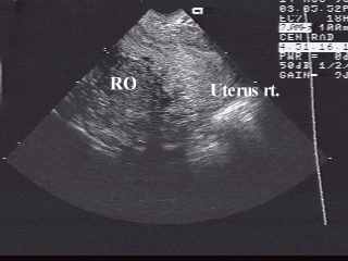
Fig.1 First TV scan.
Longitudinal sonogram of the
uterus, it markedly deviated to the right. Endometrium corresponds to secretory
phase. (LMP 25.07.98).
Right ovary is visualized posterior
to the uterus. Contains the corpus luteum.
In the region of left adnexa
the large complex mass with fluid - filled areas was indistinctly identified.
Therefore she was undertaken the transabdominal US.
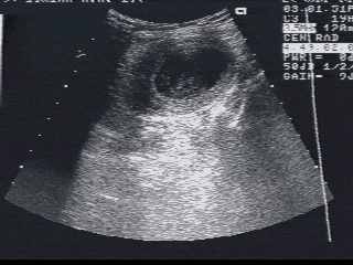
Fig.2 TA sonogram.
In the region of the left lower
quadrant the second uterus was found with distended up to 25 mm uterine
cavity filled with inhomogeneous fluid (hematometra).
It was painfull with palpation.
There was no visible communication between left cervix and vagina.
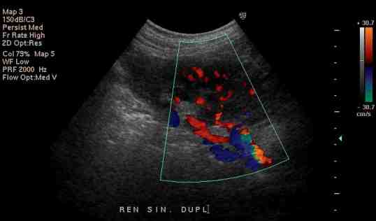
Fig.3 Sonogram of left kidney.
Color Doppler reveales an additional
artery to the low segment of left kidney.
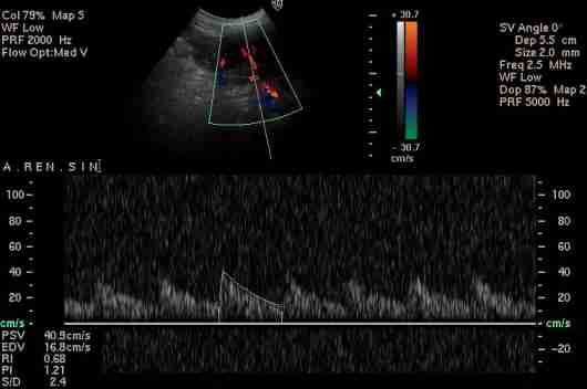
Fig.4 (a) Spectral Doppler
Waveform of a main renal artery
(a) and additional artery (b) showing the normal low resistance pattern.
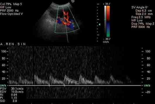
Fig.4 (b) Spectral Doppler
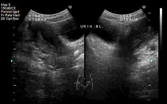
Fig.5 Follow-up TA sonography (9th day of menstrual period).
Two separate utera on both sides
of urinary bladder. Note that hematometra of the left uterus disappeared.
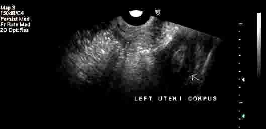
Fig.6 TV scan.
The cavity of left uterus contains
only the hyperechopic material (blood clots) and no fluid. It may suggest
a presence of communication of uterine cavity with vagina.
Any problems mail to: [email protected]