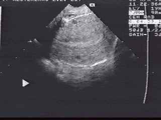


Fig.1 Longitudinal TV scan of normal
uterus.

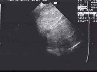
Fig.2 Pulling the probe out and turn
it to the right wall of the vagina a rounded slightly more echogenic than
myometrium lesion was founded. The myoma of vaginal wall was suggested.

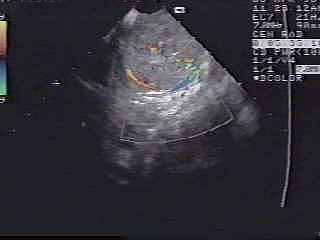
Fig.3 Color Doppler reveales internal
vessels that confirms that this lesion is not a cyst.
Pathologic diagnosis was leiomyoma of vaginal
wall.
Our Address:
Ukraine, Kyiv, 252004, Shevtchenko
blvd, 13, National Medical University, Radiology Department, Nella K. Volyk
Phone/fax: +(380)-44-2945521
![]() [email protected]
[email protected]
Any problems mail to: [email protected]