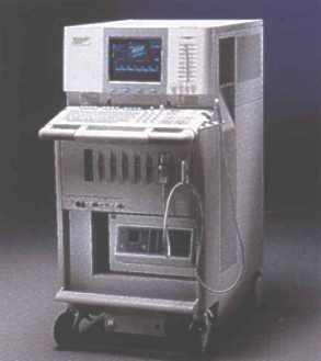
 The
Acuson 128 XP/10 system uses Acusonís unique Second Generation
Computed Sonography technology.
The main characteristics of 128XP platform are:
1. 128-electronically independent canals
provide exclusive spatial resolution, tissue contrast and image uniformity
in all field of view.
The
Acuson 128 XP/10 system uses Acusonís unique Second Generation
Computed Sonography technology.
The main characteristics of 128XP platform are:
1. 128-electronically independent canals
provide exclusive spatial resolution, tissue contrast and image uniformity
in all field of view.
2. Acustic Responce Technology (ART)
- optimises subtle signals returning from the body to account
for the proper shaping of the acustic spectrum, taking into consideration
the transduser response, tissue attenuation and user-selected parameters
such as depth, preprocessing options and operating frequency chosen throuh
MultiHertzTM.
3. Regional Expansion Selection (RES)
lets expand a portion of 2-D image and Color Doppler image and view it
in real-time. The resolution is actually increased because the system adds
more lines-both horizontal and vertical-of ultrasound data to the expanded
area.
4. Vector Array Format technology.
Vector is Acusonís trademark for its proprietary omni-steerable, omni-originating
image formation technology. Like a sector transduser, a Vector Array transduser
has a small footprint for imagingwhen access is difficult, however the
near field image width is as wide as the transduser footprint. In addition,
a Vector Array transduser produces a wider field of view at all depths
than previously possible with a sector transduser.
5. MultiHertz technology
e[tends the usefullness of a single transduser by enabling it to operate
at multiple independent imaging frequencies. This capability provides better
resolution at higher frequencies and vore gray scale and Color Doppler
penetration at lower frequencies. In addition, the lower frequencies provide
increased Color Doppler velocity scales to reduce aliasing.
6. Color Doppler Imaging Option
is an imaging technique that displays blood flow information in real-time.
The system allows to combine Color Doppler information on the 2-D image
and spectral Doppler information in a strip at the same time. Triplex function
on linear transdusers allows to simultaneously acquire and display spectral
Doppler, Color Doppler and 2-D information.
7. Color Doppler Energy (CDE) Option
lets and assigns a color to the energy generated by moving reflectors (blood
flow). CDE is virtually angle and velocity independent, displays a wide,
dynamic range of signals from blood vessels with varying flow states, ranging
from high velocity to very low velocity simultaneously without aliasing.
CDE displays perfused regions where the mean blood velocity is zero.
8. Doppler Tissues Imaging (DTI)
is a new investigational tool which presents myocardial motion abnormalities
the way Color Doppler presents blood flow abnormalities by Color encoding
the myocardium from the Doppler shift information of moving tissue. Potential
areas of application include: disorders of contractility and condactivity
and diastolic function.
DTI enables myocardial motion to be illustrated in three
ways:
-Velocity Mode-Color presentation
of mean velocity of tissue in the sample area
-Acceleration Mode-Color presentation
of the rate change of velocities in the sample
area
-Energy Mode-Color presentation
of Doppler signal energies returning from the
tissue.
The ultrasound system in
place is equipped with 10 transdusers allowing to perform the following
examinations:
1. Abdominal sonography (liver, biliary tree, pancreas, spleen, deep
abdominal vessels)
2. Urinary tract sonography (kidneys, adrenals, urinary bladder)
3. US of small parts (thyroid gland, breast, scrotum)
4. Fetal sonography (recording the examination on videotape is
available by request)
5. Gynecology US, both transabdominal and transvaginal approach
6. Neonatal Us
7. Prostate sonography
8. Carotid US
9. Transcranial US
10. Echocardiography
Special examinations:
1. US of infertile women
-
assessment of ovulation disorders
-
sonohysterography (tubal patency evaluation under the US-guidance)
2. Assessment of vertebro-basilar disorders, intervertebral discs
pathology
3. Us-guided puncture of thyroid lesions
All examinations include the evaluation of organ and lesion
perfusion.


Our Address:
Ukraine, Kyiv, 252004, Shevtchenko
blvd, 13, National Medical University, Radiology Department, Nella K. Volyk
Phone/fax: +(380)-44-2945521
 [email protected]
Any problems mail to: [email protected]
 The
Acuson 128 XP/10 system uses Acusonís unique Second Generation
Computed Sonography technology.
 The
Acuson 128 XP/10 system uses Acusonís unique Second Generation
Computed Sonography technology.